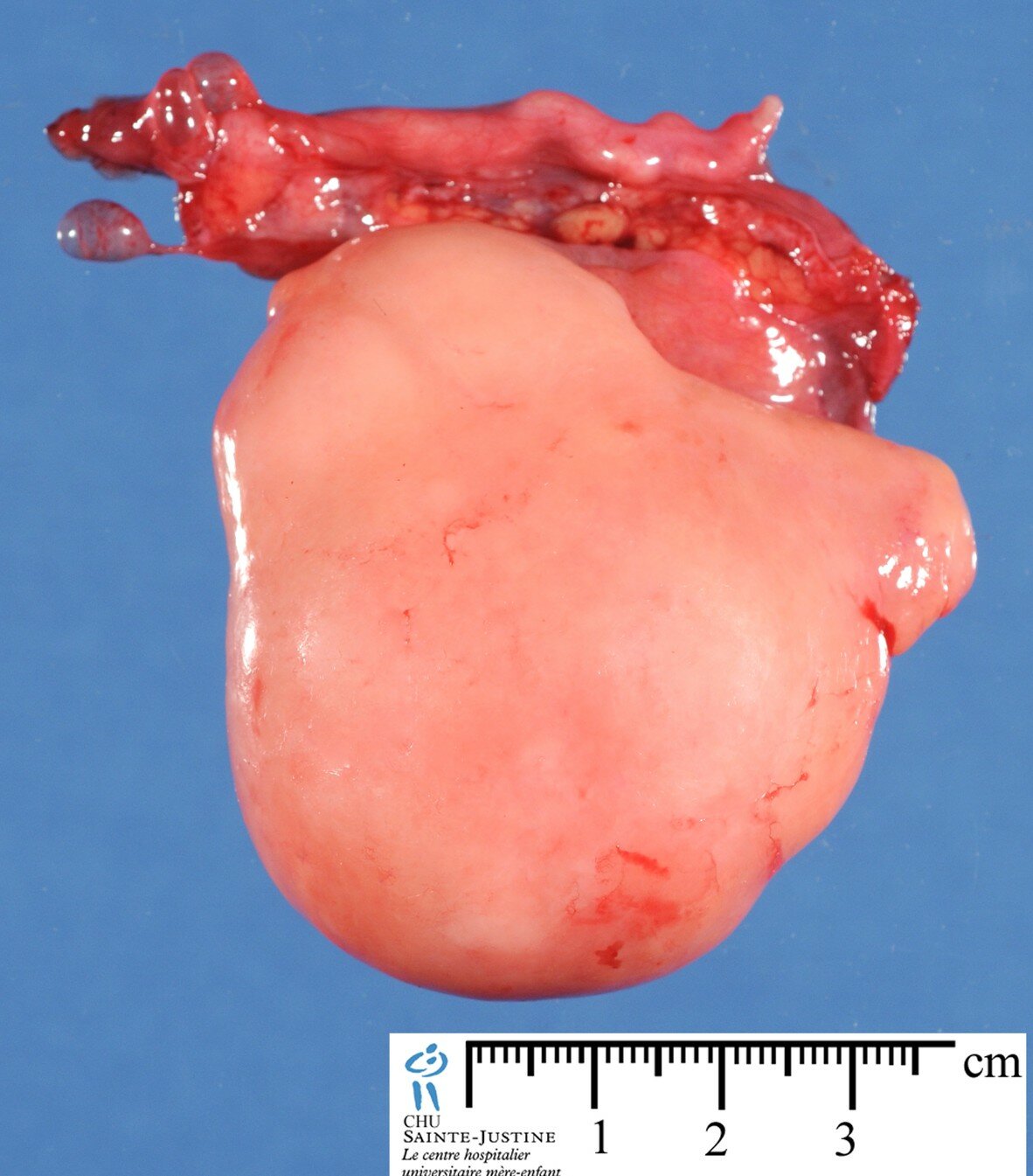
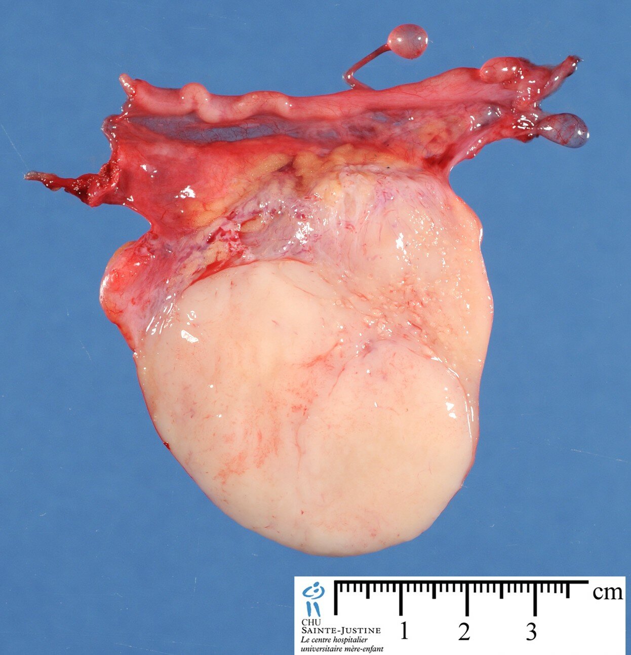
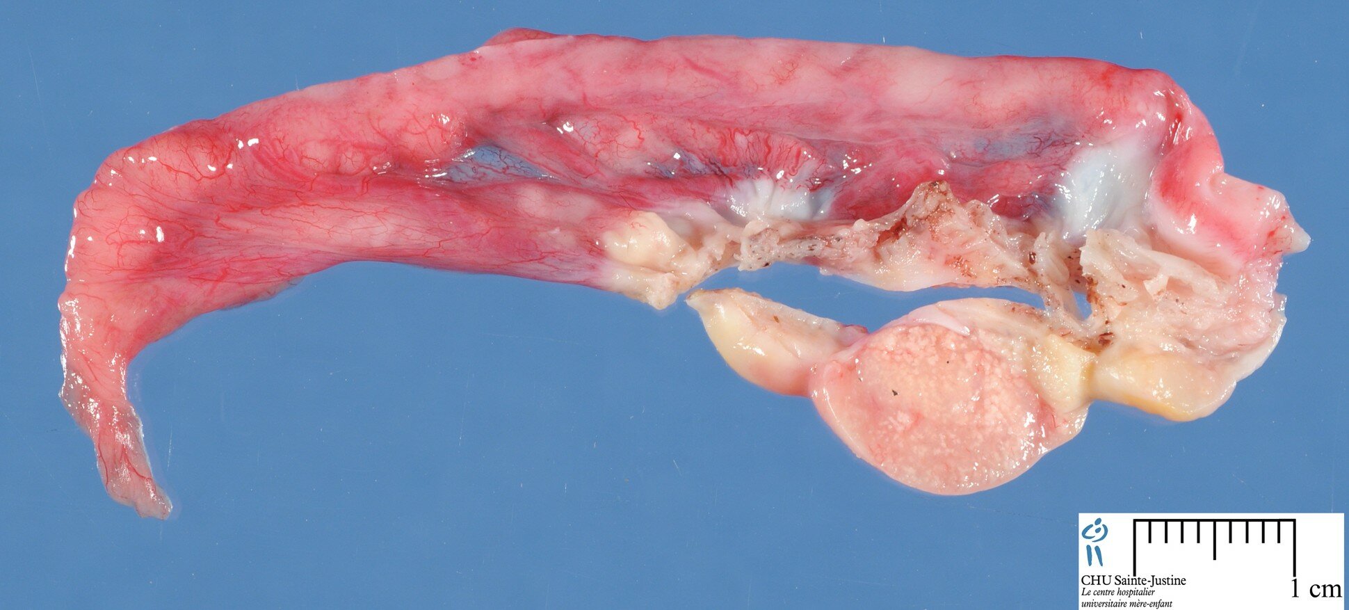
Definition: Tumor composed of a mixture of germ cell and sex-cord-stromal elements that arises almost exclusively in dysgenetic gonads.
Synopsis
 one third detected before the age of 15
frequent association with abnormalities in the secondary sex organs, with detection of a Y chromosome in over 90% of cases
bilateral in at least 38% of cases
usually develops in a phenotypic female, frequently virilized (see also gonadal dysgenesis)
may be overgrown by dysgerminoma or by other neoplastic germ cell elements
Patient usually sexually abnormal:
- commonly gonadal dysgenesis and carrying Y chromosome, i.e.:
- XY gonadal dysgenesis
- XO–XY mosaicism
- but not XX gonadal dysgenesis
estimated 25% risk of neoplasia in these dysgenetic gonads
Also documented in:
- phenotypically and chromosomally normal females, even during pregnancy
- ataxia–telangiectasia
Pathogenesis
Microscopic structures resembling gonadoblastoma found incidentally in some ovaries of normal children in association with follicular cysts may represent precursor.
Macroscopy
Usually small
Often impossible to determine nature of gonad bearing tumor
- sometimes identified as:
- a streak gonad (many become apparent only on microscopic examination)
- cryptorchid testis
- never a normal ovary
≈36% bilateral
Microscopy
germ cells, similar to those of dysgerminoma or seminoma
sex cord derivatives resembling immature sertoli or granulosa cells
stromal derivatives mimicking luteinized or Leydig cells devoid of Reinke crystals mnay be present
solid nests of inimately admixed germs cells and sex cord derivatives, the latter arranged in 3 typical patterns :
- coronal pattern at the periphery of the nests
- surrounding individual or collections of germ cells
- surrounding speces containing an eosinophilic PAS-positive material, resembling Call-Exner bodies
- sharply outlined tumor nests
- heavy calcification
Admixture of:
- primitive germ cells:
- resembling those of dysgerminoma
- sex cord–stromal cells:
- resembling morphologically and immunohistochemically immature Sertoli and granulosa cells
Steroid hormone-producing cells:
- may be present (especially after puberty)
- have capacity for steroidogenesis
additional processes :
- hyalinization
- calcification
- originating in the Call-Exner-like bodies
- when abundant may be obvious on plain abdominal radiograph
- overgrowth by a malignant germ cell component, usually dysgerminoma (50% of cases) or another malignant germ cell element (in an additional 10%)
if the nature of the gonad is identified it is either a streak or a testis (contralateral gonad : idem)
Immunohistochemistry
Hyaline material reacts strongly with anti-laminin antibodies, indicating basement membrane deposition.
Associations
30% of patients with mixed gonadal dysgenesis
less than 3% of patients with true hermaphrodism
50% of patients with 46XY pure gonadal dysgenesis
very high frequency of associated malignant germ cell tumor
dysgerminoma (malignant germ cell tumor most commonly associated with gonadoblastoma)
Differential Diagnosis
dysgerminoma
sex cord tumor with annular tubules
Prognosis
Germ cell component may overgrow stromal elements and result in formation of:
a dysgerminoma
exceptionally, another type of germ cell tumor
Much more unusually overgrowth of sex cord–stromal component may result in features of a Sertoli cell tumor.
Vaiants
bilateral gonadoblastoma
See also
gonadal tumors
intersex disorders
Links
References
A novel SRY missense mutation affecting nuclear import in a 46,XY female patient with bilateral gonadoblastoma. Hersmus R, de Leeuw BH, Stoop H, Bernard P, van Doorn HC, Brüggenwirth HT, Drop SL, Oosterhuis JW, Harley VR, Looijenga LH. Eur J Hum Genet. 2009 Dec;17(12):1642-9. PMID: 19513096
Presence of TSPY transcript and absence of transcripts of other Y chromosomal genes in a case of microscopic gonadoblastoma. Su MT, Lee IW, Kuo PL. Gynecol Oncol. 2006 Oct;103(1):357-60. PMID: 16784771
Gonadoblastoma arising in undifferentiated gonadal tissue within dysgenetic gonads. Cools M, Stoop H, Kersemaekers AM, Drop SL, Wolffenbuttel KP, Bourguignon JP, Slowikowska-Hilczer J, Kula K, Faradz SM, Oosterhuis JW, Looijenga LH. J Clin Endocrinol Metab. 2006 Jun;91(6):2404-13. PMID: 16608895
Bjersing L, Cajander S. Ultrastructure of gonadoblastoma and dysgerminoma (seminoma) in a patient with XY gonadal dysgenesis. Cancer. 1977;40:1127–1137.
Nakashima N, Nagasaka T, Fukata S, Oiwa N, Nara Y, Fukatsu T, et al. Ovarian gonadoblastoma with dysgerminoma in a woman with two normal children. Hum Pathol. 1989;20:814–816.
Pratt-Thomas HR, Cooper JM. Gonadoblastoma with tubal pregnancy. Am J Clin Pathol. 1976;65:121–125.
Goldsmith CI, Hart WR. Ataxia-telangiectasia with ovarian gonadoblastoma and contralateral dysgerminoma. Cancer. 1975;36:1838–1842.
Safneck JR, deSa DJ. Structures mimicking sex cord-stromal tumours and gonadoblastomas in the ovaries of normal infants and children. Histopathology. 1986;10:909–920.
Scully RE. Gonadoblastoma. A review of 74 cases. Cancer. 1970;25:1340–1356.
Hou-Jensen K, Kempson RL. The ultrastructure of gonadoblastoma and dysgerminoma. Hum Pathol. 1974;5:79–91.
Hussong J, Crussi FG, Chou PM. Gonadoblastoma: immunohistochemical localization of Mullerian-inhibiting substance, inhibin, WT-1, and p53. Mod Pathol. 1997;10:1101–1105.
Mackay AM, Pettigrew N, Symington T, Neville AM. Tumors of dysgenetic gonads (gonadoblastoma). Ultrastructural and steroidogenic aspects. Cancer. 1974;34:1108–1125.
Roth LM, Eglen DE. Gonadoblastoma. Immunohistochemical and ultrastructural observations. Int J Gynecol Pathol. 1989;8:72–81.
Hart WR, Burkons DM. Germ cell neoplasms arising in gonadoblastomas. Cancer. 1979;43:669–678.
Govan ADT, Woodcock AS, Gowing NFC, Langley FA, Neville AM, Anderson MC. A clinico-pathological study of gonadoblastoma. Br J Obstet Gynecol. 1977;84:222–228.