



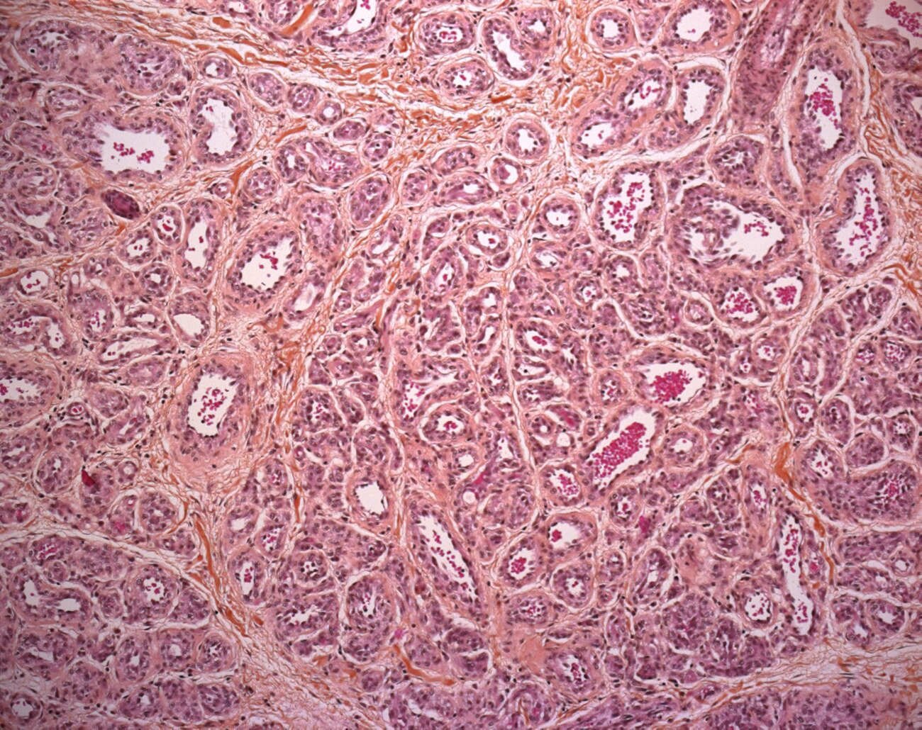
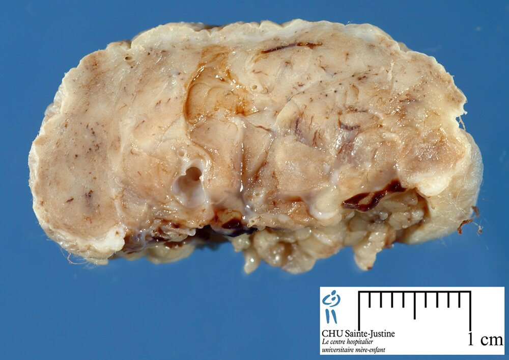
CLINICAL: The typical infantile hemangioma appears postnataly, grows quickly, and regresses gradually in the first few years of life. Approximately one third of IH are present at birth.
Infantile hemangiomas progress through 3 stages: proliferative, involuting, and involuted. The proliferative phase is characterized by clinical growth. Once hemangiomas reach their maximum size, they begin to regress or involute. Histologically, this stage is characterized by endothelial apoptosis. Finally, the involuted stage of the hemangioma occurs when the original lesion is replaced by a connective tissue remnant. In contrast to hemangiomas, vascular malformations do not involute but continue to enlarge as the patient grows.
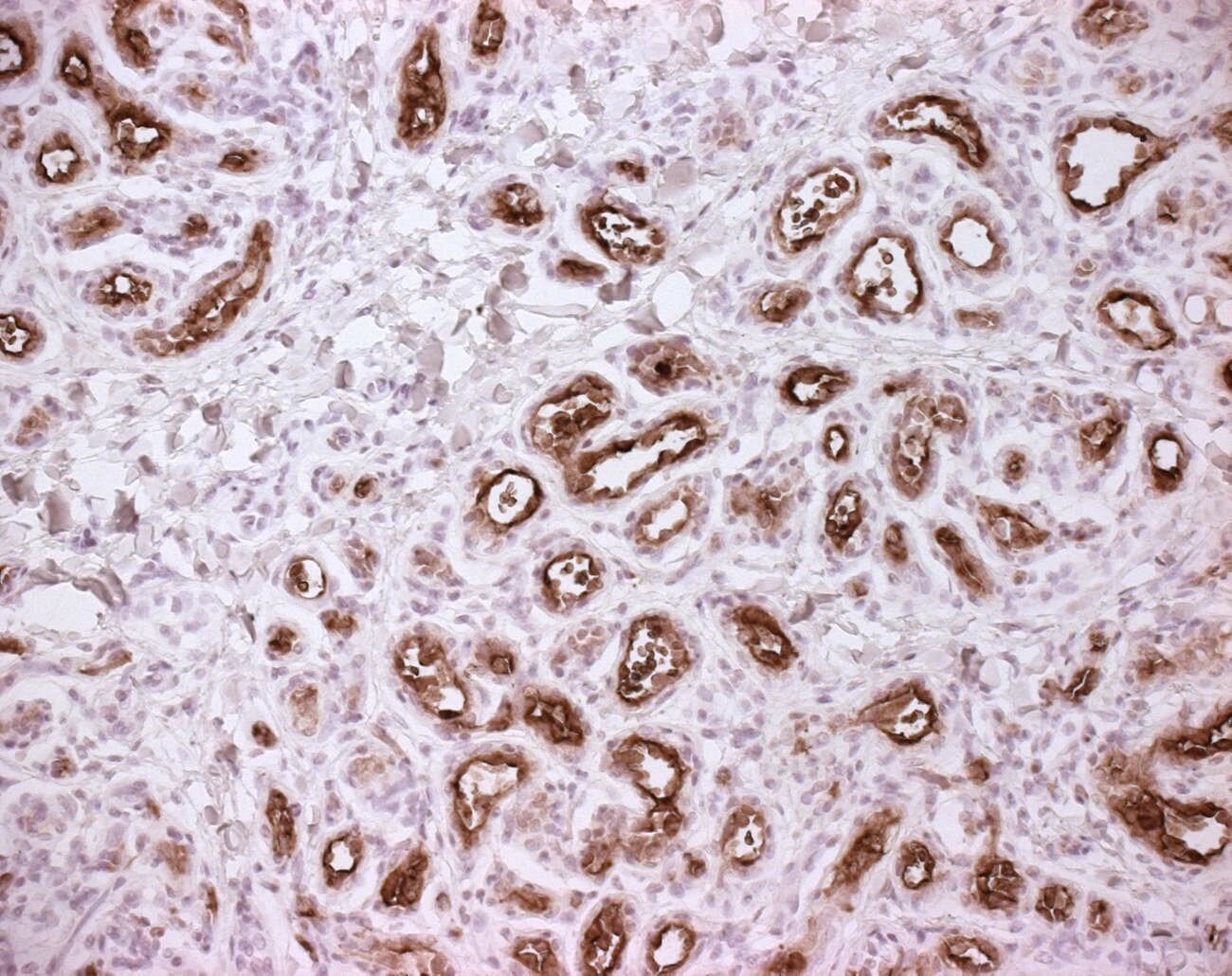
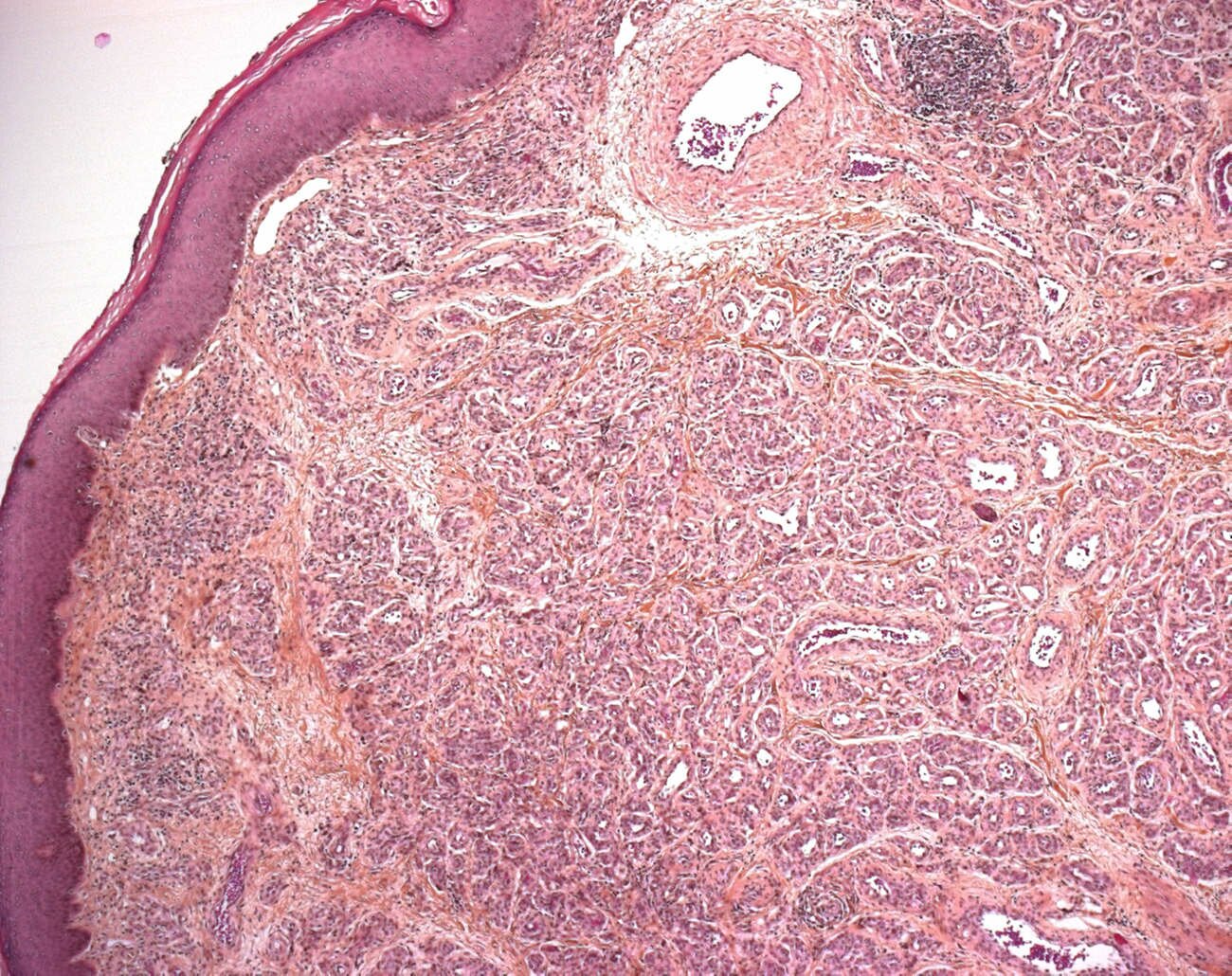
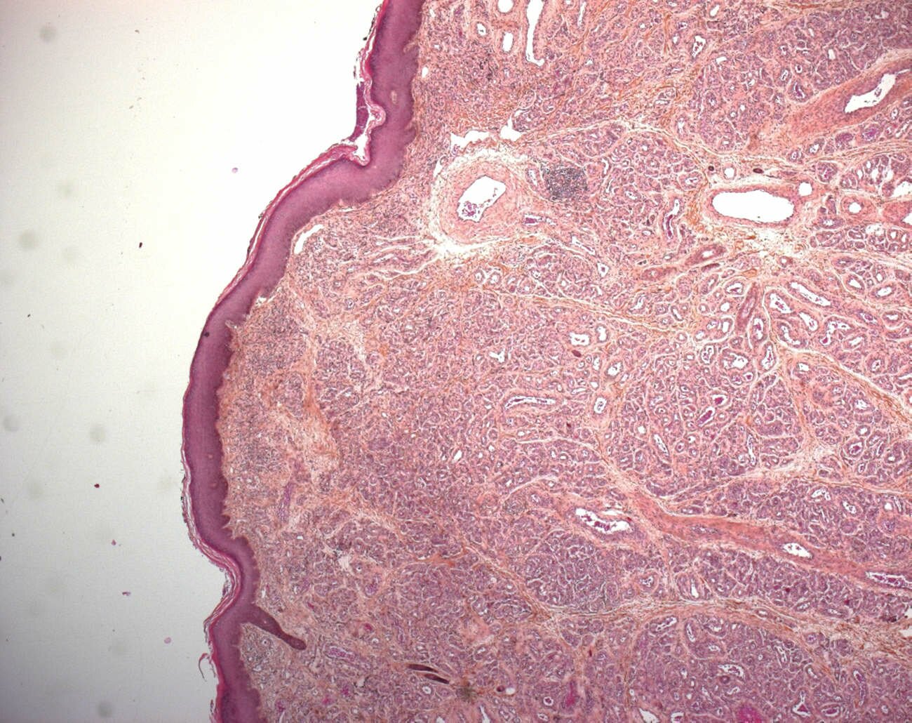
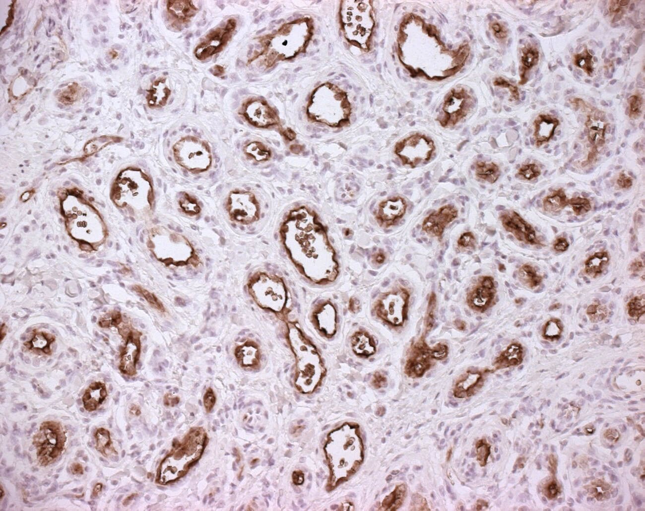
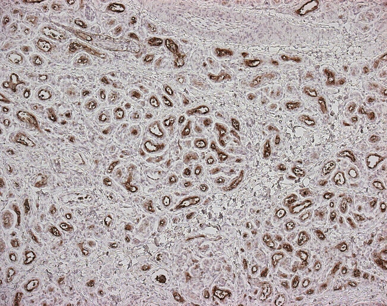
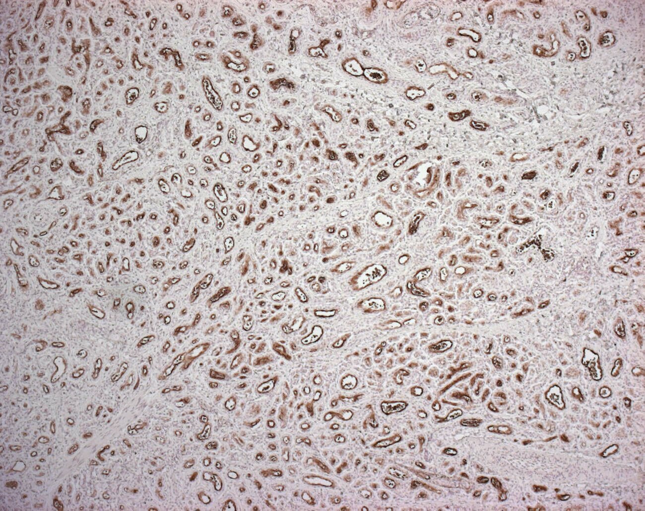
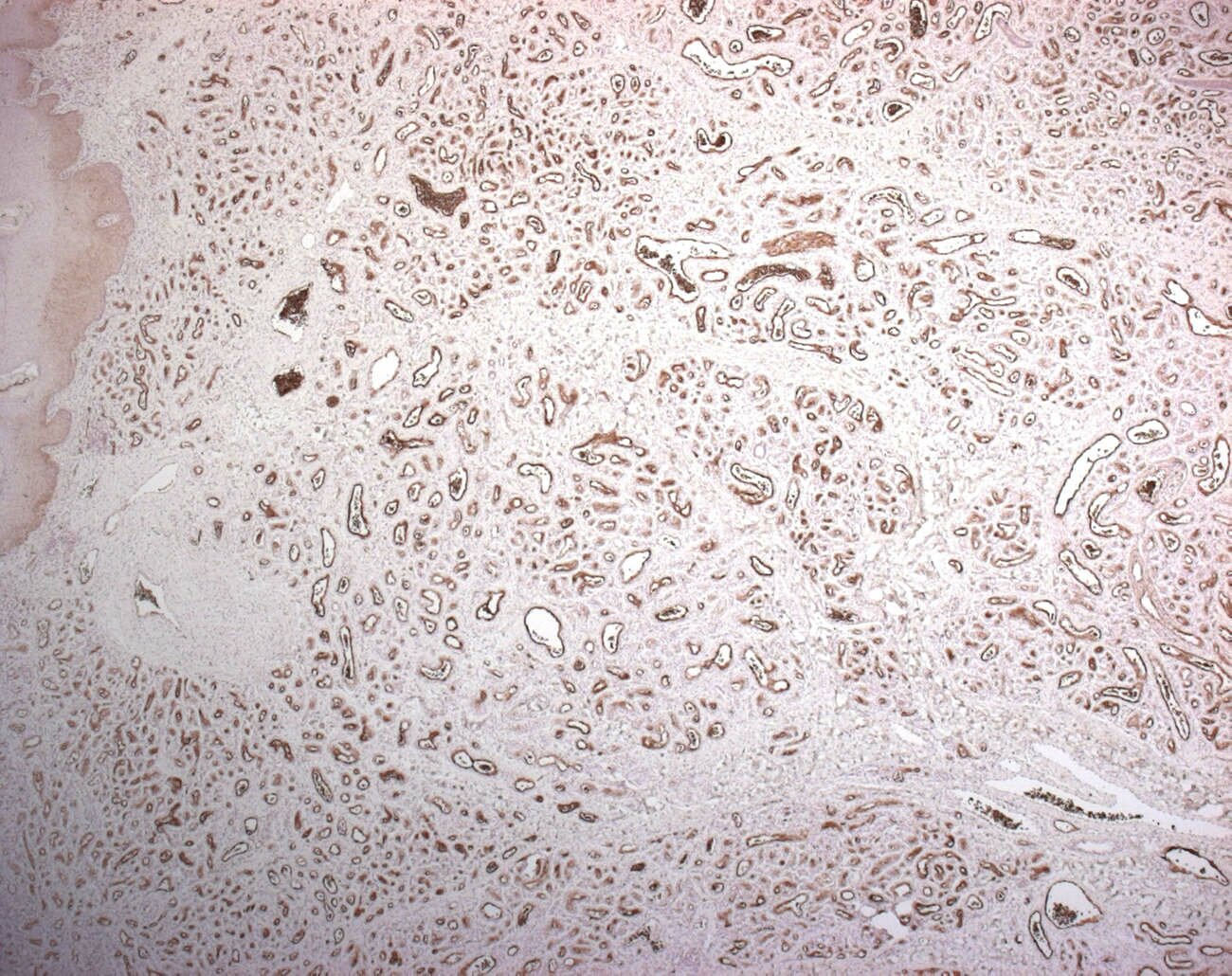
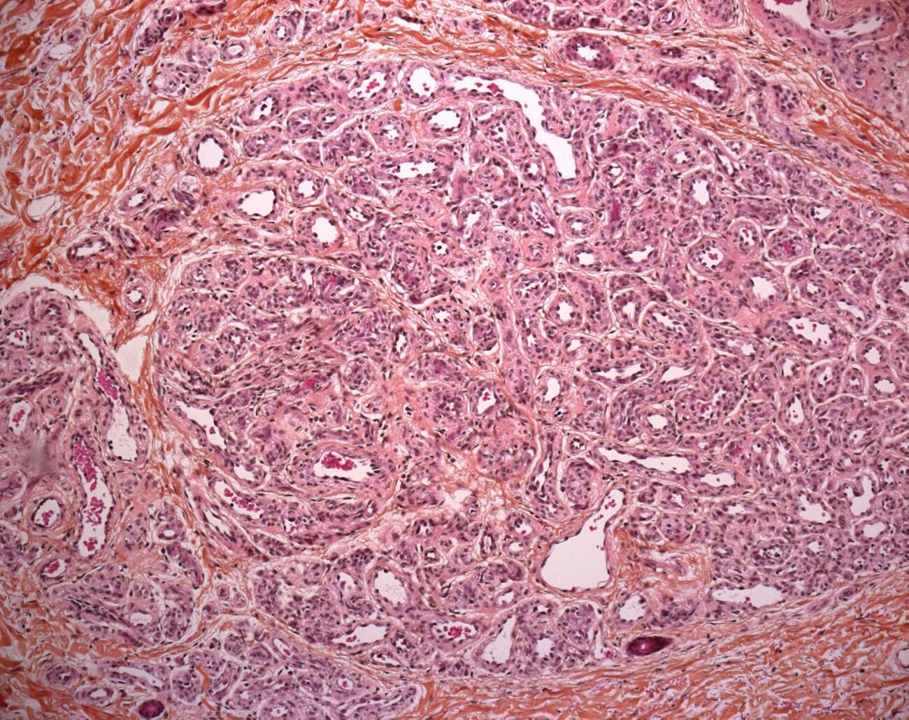
HISTOPATHOLOGY: these tumors are highly cellular, involving the dermis, often with extension into the subcutis. The tumor has a lobular architecture and plump endothelial cells. The latter cells express glut-1 antigens throughout the life of this tumor.
Synopsis
 lobular architecture
Developmental phases:
proliferative phase
involutive phase
end-stage
Localization
cutaneous infantile hemangioma
extra-cutaneous GLUT1+ infantile hemangioma
- hepatic infantile hemangioma (15509684)
- mammary infantile haemangioma (15509684)
- salivary infantile haemangioma (15509684)
- placental infantile haemangioma (15509684)
Variants
multiple infantile hemangioma (16880251)
reticular infantile hemangioma (17845155)
Associations
type 2 infantile hepatic hemangioendothelioma (16880251)
Differential diagnosis
congenital hemangiomas
vascular malformations
Immunochemsitry
WT1+
GLUT1+ (16230568)
| Tumors | GLUT1 | WT1 |
| infantile hemangioma | + | + |
| vascular malformation | - | - |
Predisposition
Beckwith-Wiedemann syndrome (15509684)
familial predisposition at locus 5q31-33
Molecular biology
monoclonality
RETURN TO: vascular tumors
References
Lawley LP, Cerimele F, Weiss SW, North P, Cohen C, Kozakewich HP, Mulliken JB, Arbiser JL. Expression of Wilms tumor 1 gene distinguishes vascular malformations from proliferative endothelial lesions. Arch Dermatol. 2005 Oct;141(10):1297-300. PMID: 16230568
Timar J, Meszaros L, Orosz Z, Albini A, Raso E. WT1 expression in angiogenic tumours of the skin. Histopathology. 2005 Jul;47(1):67-73. PMID: 15982325
Dadras SS, North PE, Bertoncini J, Mihm MC, Detmar M. Infantile hemangiomas are arrested in an early developmental vascular differentiation state. Mod Pathol. 2004 Sep;17(9):1068-79. PMID: 15143338