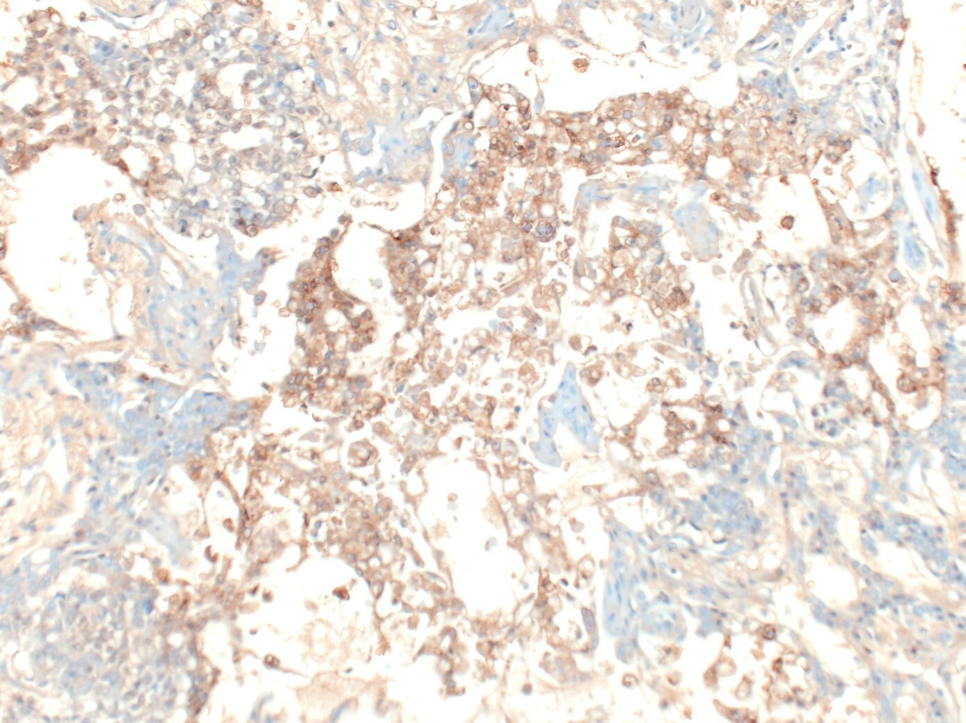

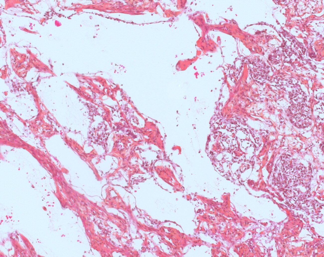
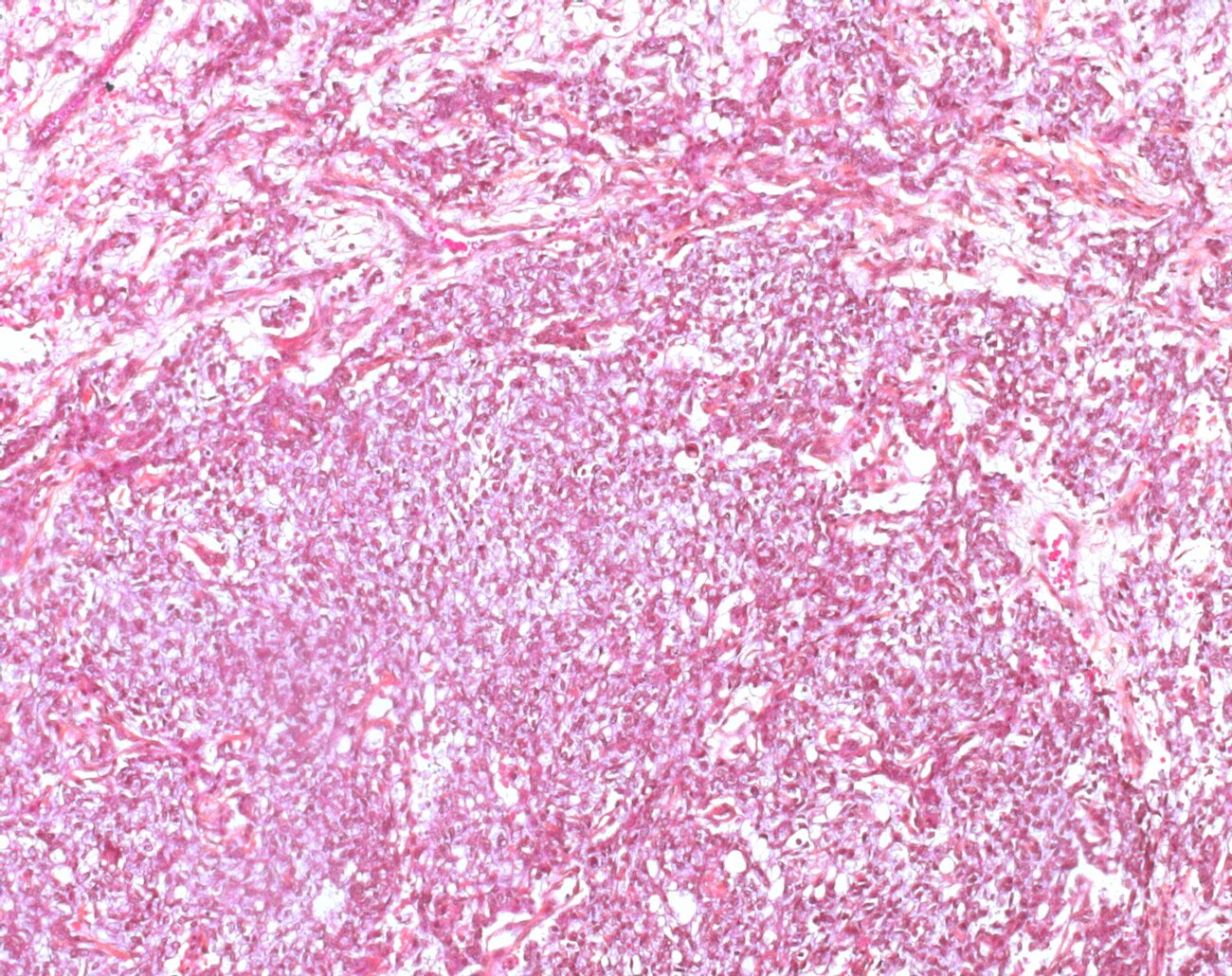
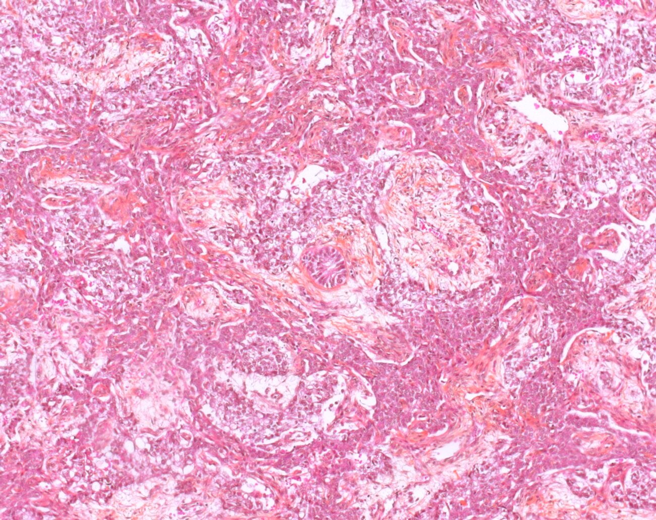
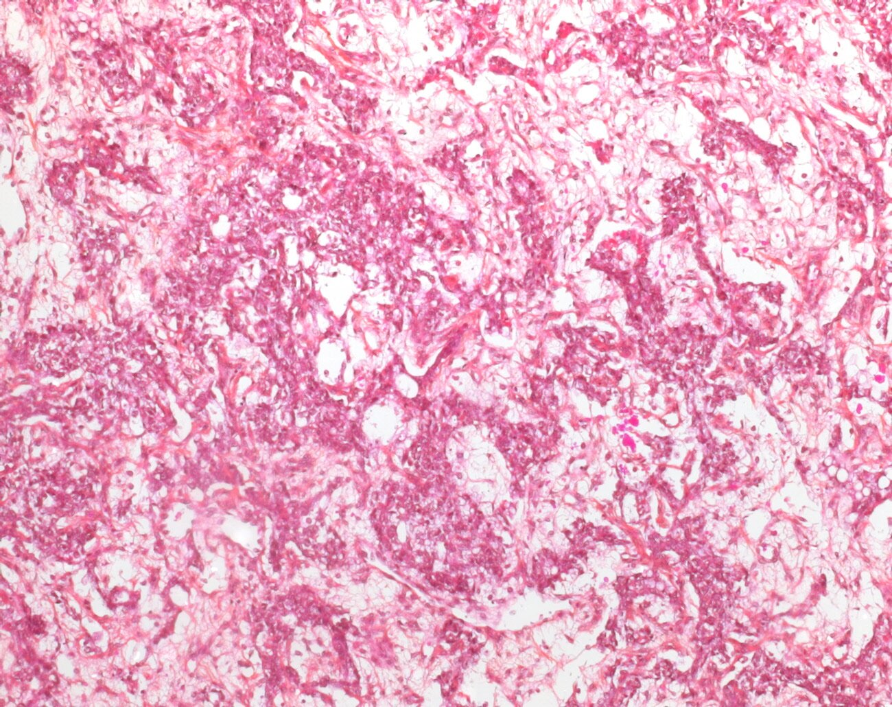
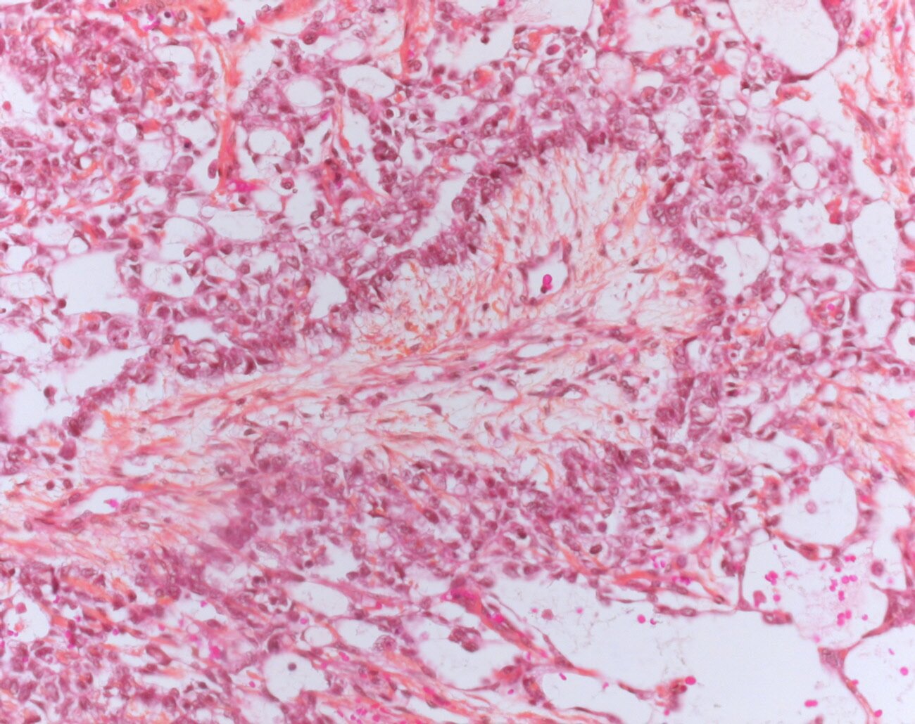
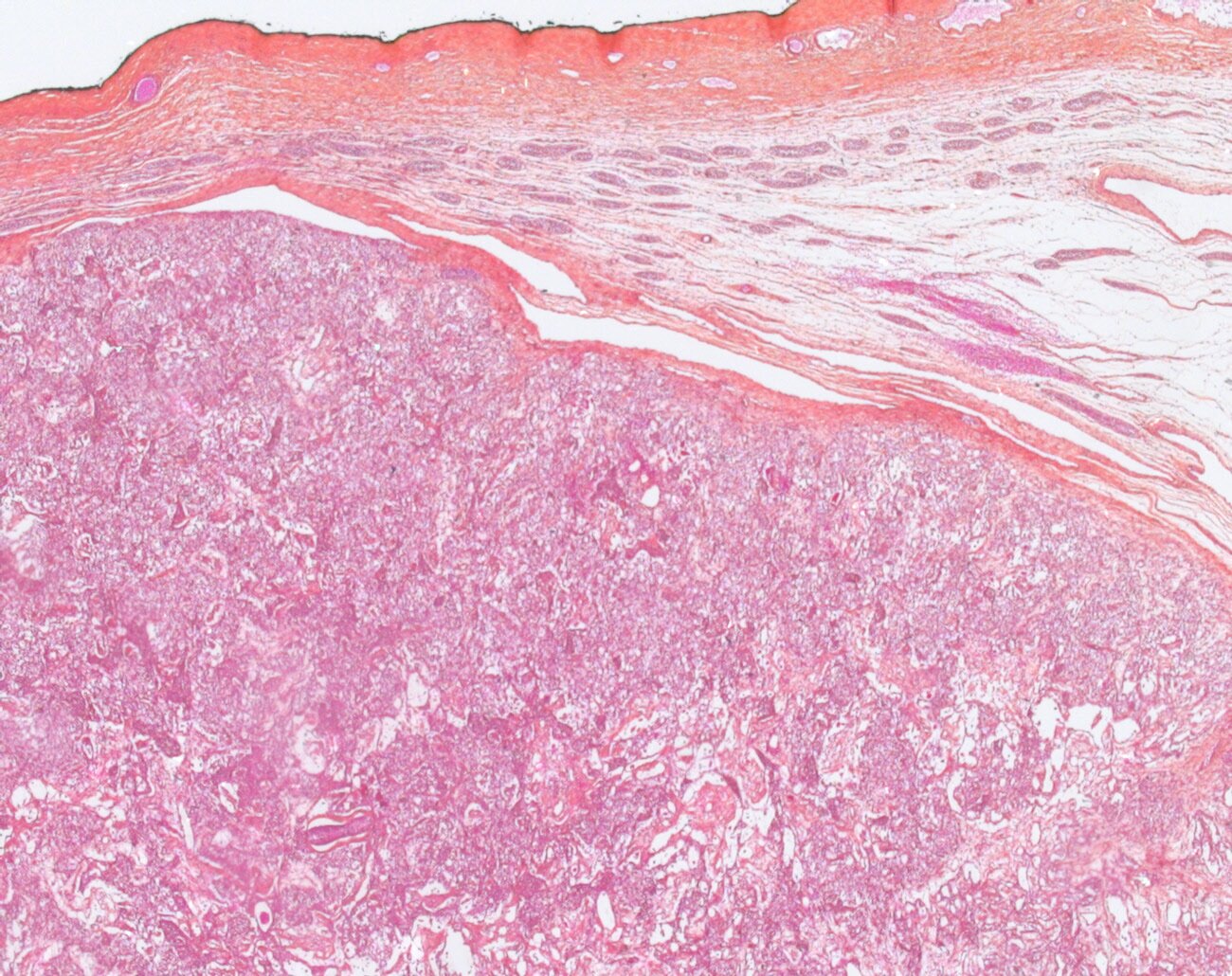

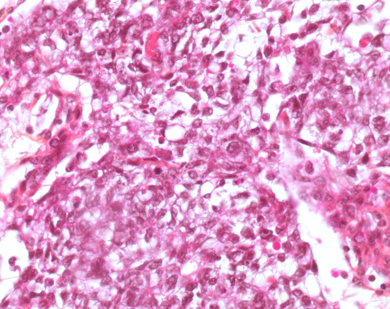

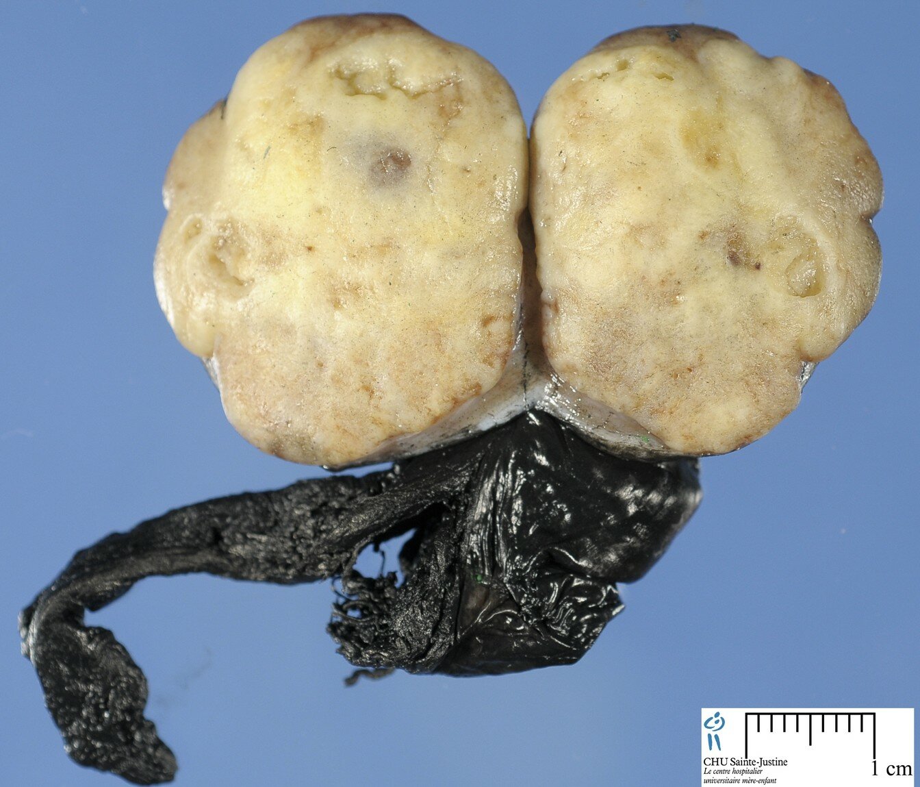


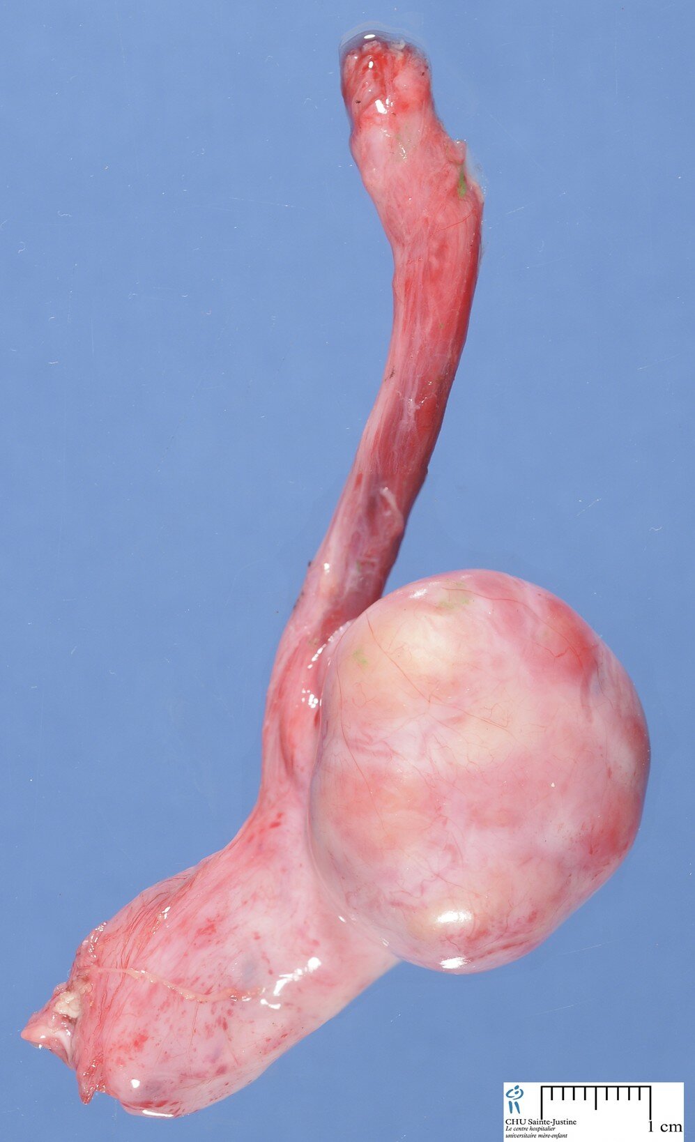

Definition: The yolk sac tumor is a malignant germ cell neoplasm that recapitulates the primary embryonic yolk sac tissue (endodermal sinus).
YST is the most common malignant testicular germ cell tumor of pediatric population (60 to 70%).
Intratubular neoplasia (ITGCN) is absent.
Epidemiology
 Age: six months to five years, peak in boys of age less than 2 years
Pathogenesis
Hypermethylation of the RUNX3 gene promoter.
Pediatric cases express GATA-4, a transcription factor that regulates differentiation and function of murine yolk sac endoderm
Treatment
1. Surgical Management: Radical Orchiectomy with high ligation of the spermatic cord with Retroperitoneal Lymph Node Dissection
- Indications
2. Chemotherapy : Platinum-based regimens seems to apply to childhood germ cell Prognosis Excellent (5 year survival 90% even in advanced cases)
Variants
prepubertal yolk sac tumor
- Isochrome 12p is not seen
- Euploid or tetraploid
- Recurrent nonrandom chromosomal abnormalities: 1p deletion, 3p duplication, 6q deletion
- p53 gene mutations absent.
- Equal incidence of lymphatic and hematogenous spread for metastases.
- Presents as stage I disease (85%)
adult yolk sac tumor
- presents along with other germ cell tumor components.
- Isochrome 12p positive
- Intratubular neoplasia is present.
- Usually aneuploid p53 gene mutations.
- Lymphatic route is primarily spread for metastases.
- Often presents as higher stage disease
Microscopical synopsis
microcystic pattern
- interconnecting cords and ribbons of tumor cells are surrounded by abundant myxoid stroma
- intracellular vacuoles
- vesicular nuclei with punctate nucleoli
- often coexists with other architectural patterns
myxomatous pattern
- neoplastic stellate cells
- spindle cells
- epithelioid cells
- abundant myxoid stroma
- many of these cells are pluripotential and can form skeletal muscle, cartilage, and bone (such areas should not be confused with teratoma)
endodermal sinus pattern
- Schiller-Duvall body: central vessel surrounded by tumor cells in a cystic space often lined by flattened tumor cells
hepatoid pattern (20%)
- sheets of polygonal cells with abundant eosinophilic cytoplasm
- hyaline globules
- bile canaliculi
solid pattern
- sheets of uniform tumor cells with clear or pale pink cytoplasm
- differential diagnosis: seminoma (it lacks the fibrous septa with lymphoid infiltrate seen in seminoma)
Cytogenetics
deletion of chromosome 1p36 (9162192)
deletion of distal regions of chromosome 6q
Molecular biology
LOH of 6q (72%) (11753688)
LOH of 1p (45%) (11753688)
Differential diagnosis
embryonal Carcinoma
mature teratoma and immature teratoma
Sertoli cell tumor
seminoma
Case records
[Case HP #14827->14827]